TRACE: Temporal Radiology with Anatomical Change Explanation for Grounded X-Ray Report Generation
Abstract
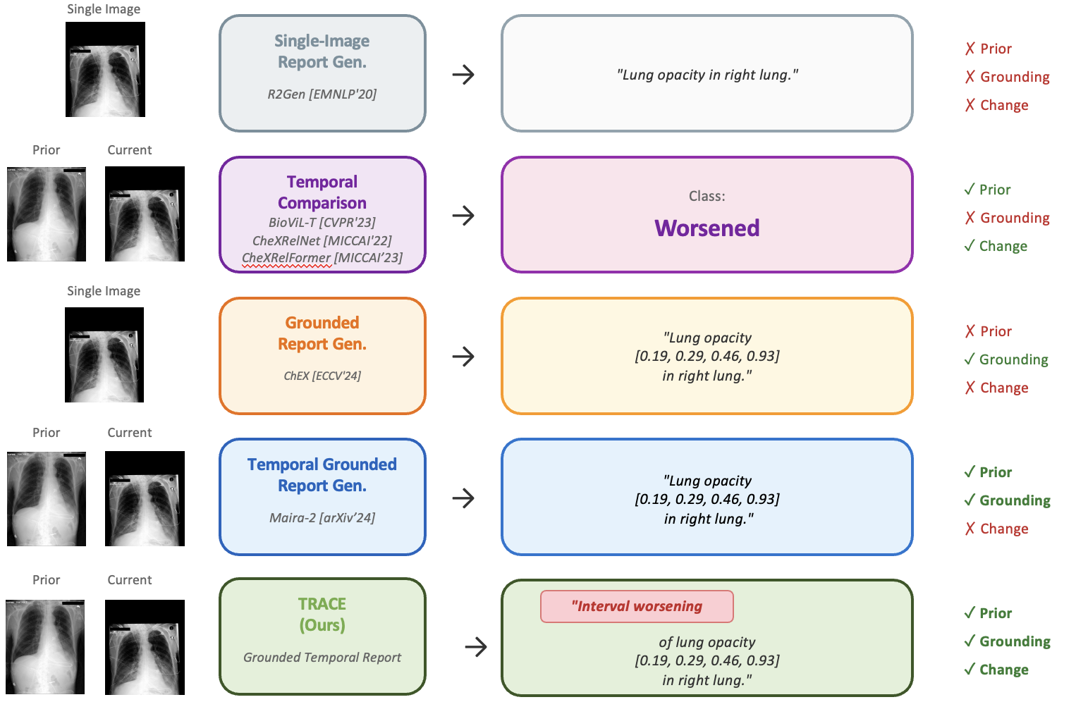
Temporal comparison of chest X-rays is fundamental to clinical radiology, enabling detection of disease progression, treatment response, and new findings. While vision-language models have advanced single-image report generation and visual grounding, no existing method combines these capabilities for temporal change detection. We introduce Temporal Radiology with Anatomical Change Explanation (TRACE), the first model that jointly performs temporal comparison, change classification, and spatial localization. Given a prior and current chest X-ray, TRACE generates natural language descriptions of interval changes (worsened, improved, stable) while grounding each finding with bounding box coordinates. TRACE demonstrates effective spatial localization with over 90% grounding accuracy, establishing a foundation for this challenging new task. Our ablation study uncovers an emergent capability: change detection arises only when temporal comparison and spatial grounding are jointly learned, as neither alone enables meaningful change detection. This finding suggests that grounding provides a spatial attention mechanism essential for temporal reasoning.
What the failure looks like
The emergent capability is easiest to see in how the ablated model breaks. Give it both images but remove grounding supervision and it does not degrade gracefully: it collapses to predicting "stable" for every single sample, scoring 0% recall on both worsening and improvement. The 43.8% accuracy it reports is just the majority class rate, not evidence of any learned comparison.
Example TRACE Output:
Key Contributions
- We introduce grounded temporal change detection, a new task combining temporal comparison, change classification, and spatial localization in a unified framework.
- We discover an emergent capability: change detection only arises when both temporal comparison and spatial grounding are combined. Without grounding supervision, models fail to detect changes even with access to both images.
- We construct a large-scale dataset with grounded temporal annotations from MIMIC-CXR and Chest ImaGenome (79,202 training, 11,464 validation, 22,553 test samples).
Method
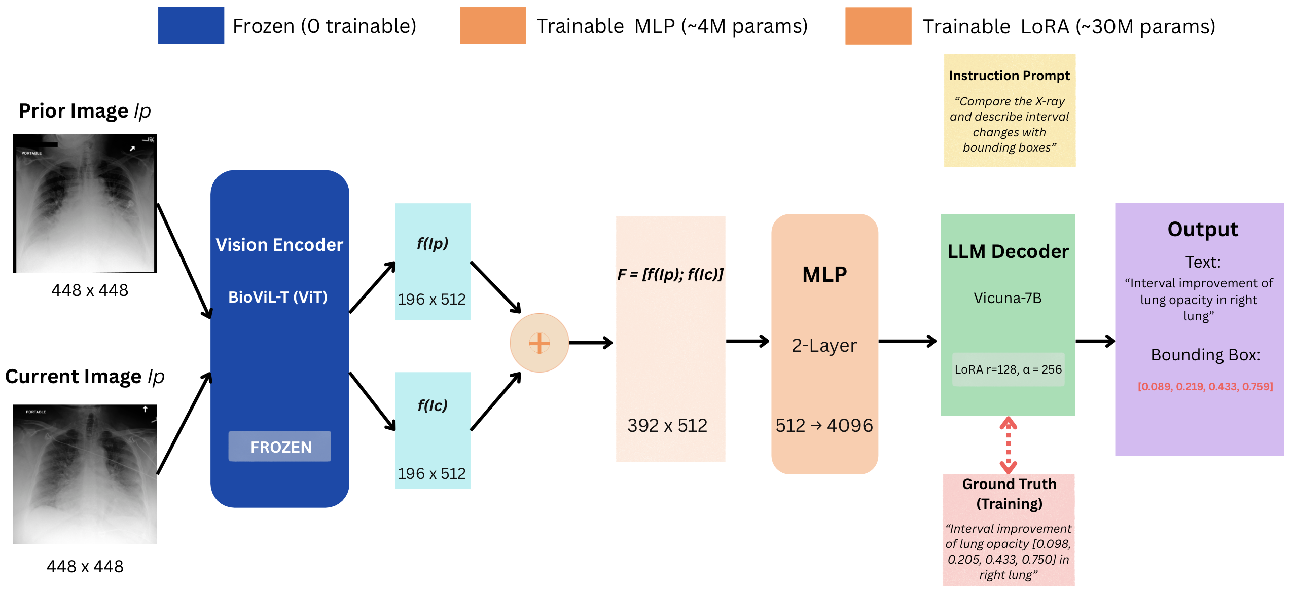
Figure: TRACE architecture for grounded temporal change detection. Given a prior and current chest X-ray, the model generates natural language descriptions of interval changes while localizing each finding with bounding box coordinates.
Temporal Input
Takes both prior and current chest X-ray images to enable comparison of disease progression over time.
Spatial Grounding
Localizes each finding with bounding box coordinates, enabling precise identification of where changes occur.
Change Classification
Classifies findings as worsened, improved, or stable to track disease progression and treatment response.
Report Generation
Generates natural language descriptions combining temporal changes with spatial localization.
Results
Main Results
TRACE performance on the test set (22,553 samples):
| Category | Metric | Value |
|---|---|---|
| NLG Metrics | BLEU-4 | 0.260 |
| METEOR | 0.438 | |
| ROUGE-L | 0.494 | |
| Clinical Efficacy | RadGraph F1 | 0.406 |
| CheXbert F1 | 0.432 | |
| Grounding | Mean IoU | 0.772 |
| IoU > 0.5 | 90.2% | |
| Change Detection | Accuracy | 48.0% |
Comparison with Prior Methods
| Method | Temporal Acc. | Grounding | Report Gen |
|---|---|---|---|
| CheXRelNet | 46.8% | ✗ | ✗ |
| CheXRelFormer | 49.3% | ✗ | ✗ |
| TRACE (Ours) | 48.0% | ✓ (90.2%) | ✓ |
TRACE achieves comparable temporal accuracy while providing two additional capabilities: spatial grounding and full natural language report generation.
Per-Class Change Detection
| Class | Precision | Recall | F1 | Support |
|---|---|---|---|---|
| Worsening | 0.485 | 0.375 | 0.423 | 7,787 |
| Improvement | 0.382 | 0.263 | 0.311 | 4,888 |
| Stable | 0.505 | 0.674 | 0.578 | 9,878 |
| Macro Average | 0.457 | 0.437 | 0.437 | 22,553 |
Per-Anatomy Analysis
| Anatomy | Accuracy | Support |
|---|---|---|
| Mediastinum | 75.5% | 339 |
| Cardiac Silhouette | 68.8% | 1,868 |
| Right Hilar | 61.7% | 60 |
| Right Lung | 45.3% | 17,343 |
| Left Lung | 44.7% | 2,549 |
Top 5 regions shown. A further 394 samples from other anatomical regions are omitted for brevity, which is why the support column sums to 22,159 rather than the full 22,553.
Cardiac changes are substantially easier to detect than pulmonary changes, as cardiac changes involve distinct boundary shifts while pulmonary changes are often more diffuse and subtle.
Qualitative Results
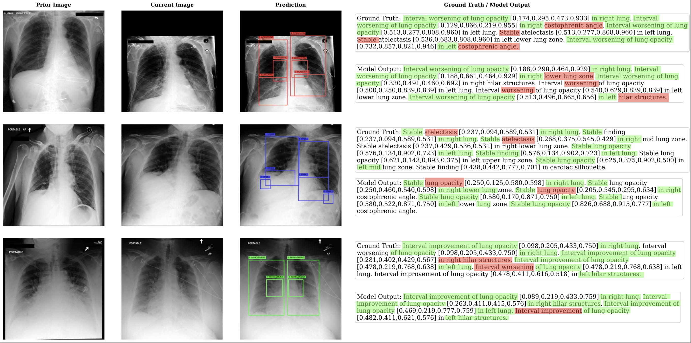
Figure: Qualitative results of TRACE on three representative cases: worsening, stable, and improvement. Each row shows the prior image, current image, model prediction with bounding box overlay, and the corresponding ground truth and model output text. The model successfully identifies temporal changes and localizes the affected anatomical regions with accurate bounding box coordinates.
Ablation Study
We conduct ablation experiments to understand the contribution of temporal input and grounding supervision:
| Model | Prior Image | Grounding | BLEU-4 | Change Accuracy |
|---|---|---|---|---|
| TRACE (Full) | ✓ | ✓ | 0.260 | 48.0% |
| Single-Image | ✗ | ✓ | 0.051 | 43.8%† |
| No-Grounding | ✓ | ✗ | 0.240 | 43.8%† |
† Predicts "stable" for all samples (0% recall on worsening and improvement). Removing the prior image leaves grounding accuracy relatively intact at 83.7%, so single-image localization is learnable without temporal context. The reverse is not true.
Critical Finding
Despite having access to both prior and current images, the no-grounding variant fails at change detection, achieving only 43.8% accuracy by predicting "stable" for all samples. This suggests that grounding supervision provides a critical inductive bias for temporal comparison. Without explicit spatial localization, the model cannot effectively compare corresponding anatomical regions across time points.
Language Model Comparison
Comparison of Vicuna-7B and Mistral-7B backbones, with the vision encoder held fixed, reveals distinct clinical behaviors:
| LLM Backbone | Accuracy | Worsening Recall | Improvement Recall | Stable Recall | IoU > 0.5 |
|---|---|---|---|---|---|
| Vicuna-7B | 48.0% | 37.5% | 26.3% | 67.4% | 90.2% |
| Mistral-7B | 45.0% | 73.6% | 57.9% | 15.7% | 54.5% |
Clinical Implications: Vicuna-7B achieves higher accuracy through conservative prediction (high specificity, lower sensitivity), suitable for screening applications where false alarms are costly. Mistral-7B exhibits aggressive change detection (high sensitivity, lower specificity), better suited for high-risk monitoring where missing actual changes is unacceptable.
BibTeX
@InProceedings{aranya2027trace,
  author    = {Aranya, OFM Riaz Rahman and Desai, Kevin},
  title     = {TRACE: Temporal Radiology with Anatomical Change Explanation for Grounded X-Ray Report Generation},
  booktitle = {Pattern Recognition (ICPR)},
  publisher = {Springer Nature Switzerland},
  address   = {Cham},
  pages     = {95--108},
  year      = {2027},
  doi       = {10.1007/978-3-032-31933-3_7},
  isbn      = {978-3-032-31933-3}
}
Acknowledgments
This work builds upon RaDialog, LLaVA, and BioViL-T. We use the MIMIC-CXR and Chest ImaGenome datasets.